Re-post from Nov 2019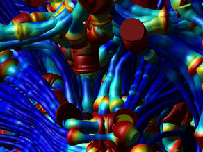
(how time flies!...)
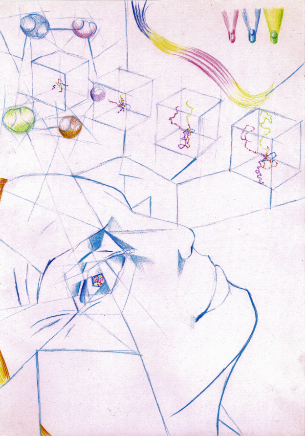
I have been creating pieces in the genre known as “artsci” since 2012, when I first met Professor Derek Jones, Director of the Cardiff University Brain Research Imaging Center (CUBRIC) at a local art exhibition where his team present their visual data – images of the brain – as art. This inspired me to bring scientific ideas to life through my painting and illustrations. For the past six years I have been commissioned to create and exhibit a range of artsci pieces that explore subjects ranging from neuroscience to magnetism and astronomy. I find the beauty in the data irresistible. Memory Sketches of Conversations at CUBRIC in 2012...
For all of my commissions, I have worked closely with scientists to understand their research and incorporate their ideas into my work. These collaborations have had a profound influence on my perspectives of how we experience our existence.
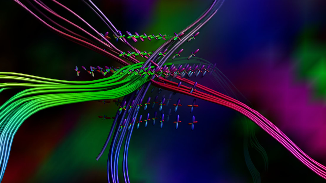
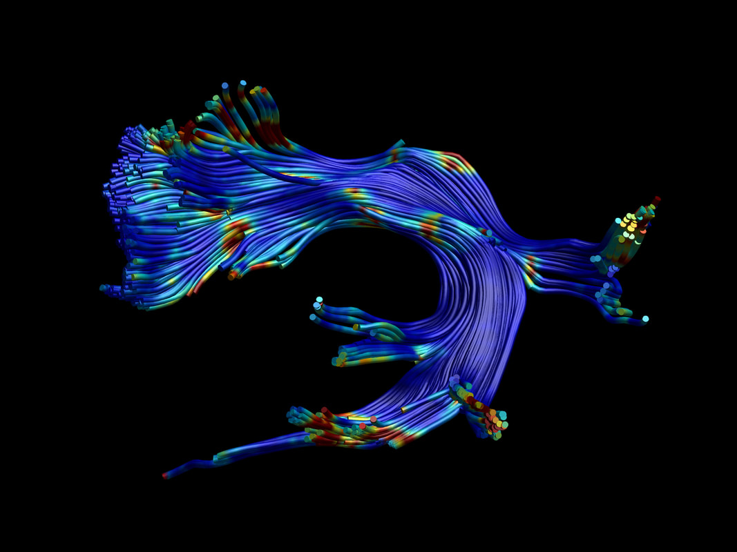
I have been lucky enough to have a glimpse of the potential of the research at CUBRIC. Inspired by their incredible images of the brain, with every new piece of knowledge I gain from my collaborators, I find myself meditating on the composition of thoughts themselves. Such as the “spin echo” of a hydrogen atom’s proton as it vibrates inside a magnetic resonance imaging (MRi) machine. Or how through “tractography” we can map the nerve cell tracts in the brain by measuring the movement of water molecules as they travel along neurons, pushed by magnetic fields. In a way, it feels like we can see the shape of our minds, and I find it incredible and very inspiring. A selection of visual data from CUBRIC...
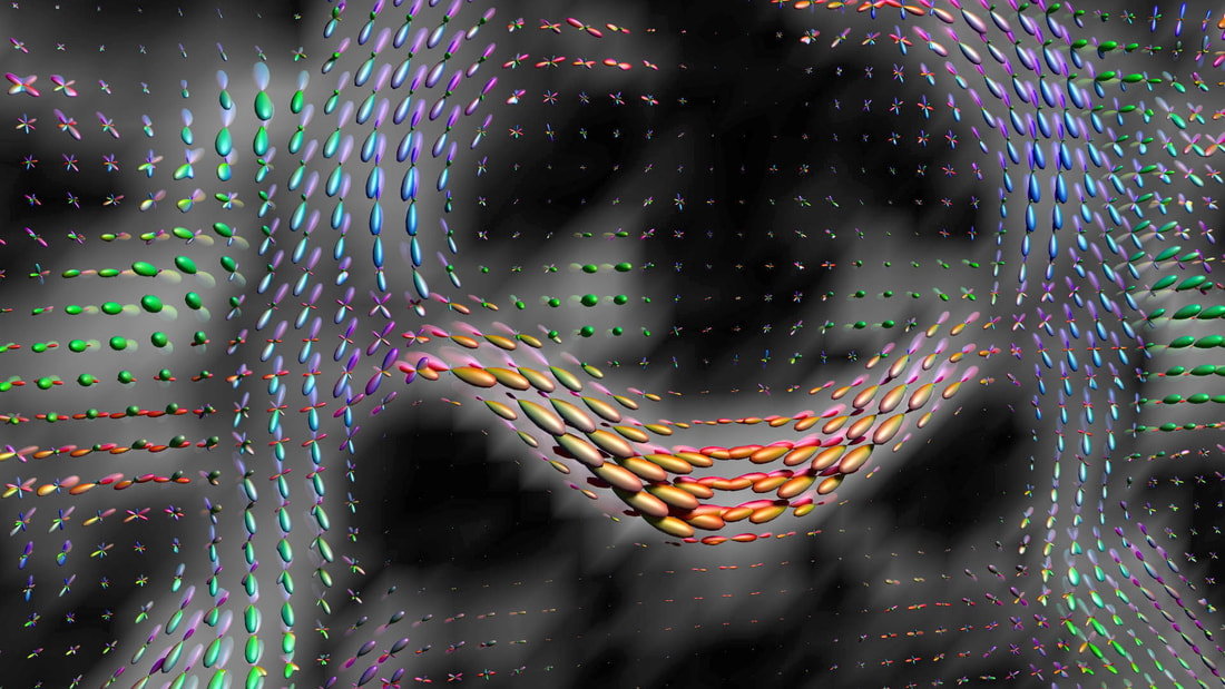
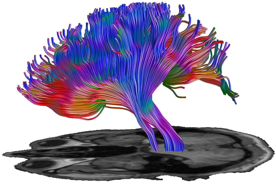
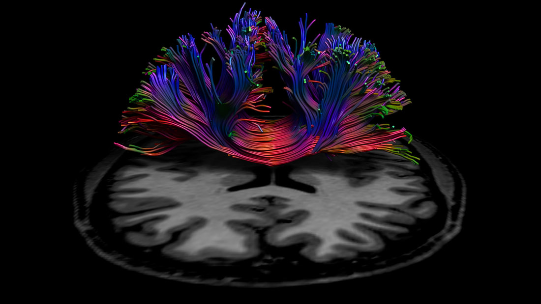
Provide by Alex Leemans of
PROcessing & VIsualization in Diffusion Imaging (PROVIDI) Lab http://www.providi-lab.org/image-gallery.html
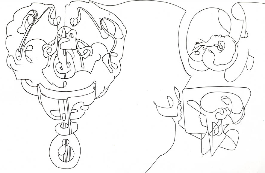
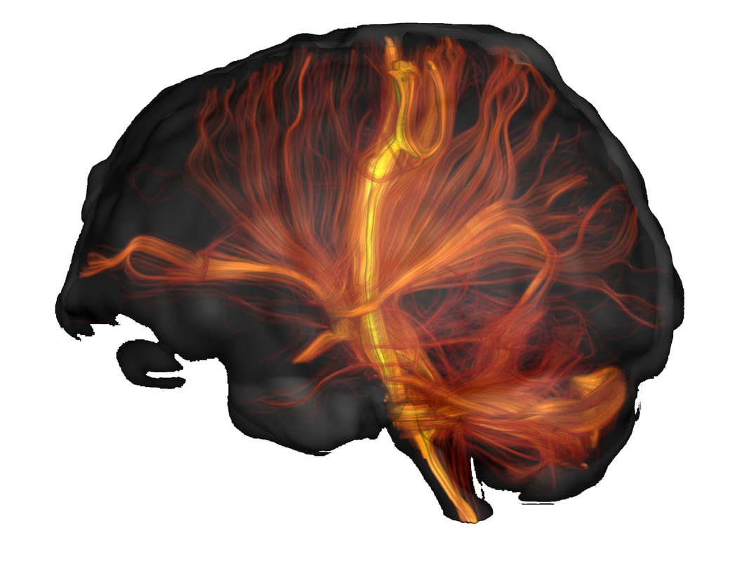
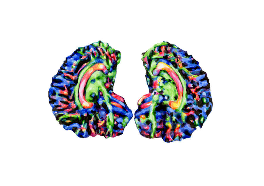
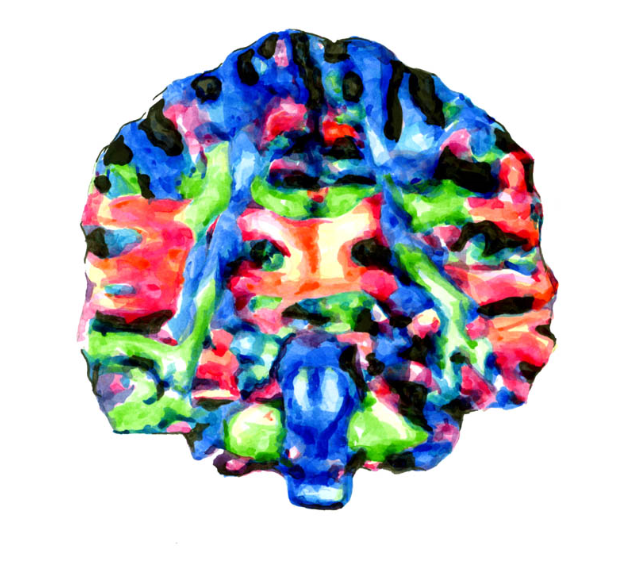
Research in the macro scale and the micro scale into the anatomy of the brain lies on spectrum so vast, it seems the deeper one delves into the structure of the mind, the further down the axons we fly into a profusion of probabilities. It blows my mind.
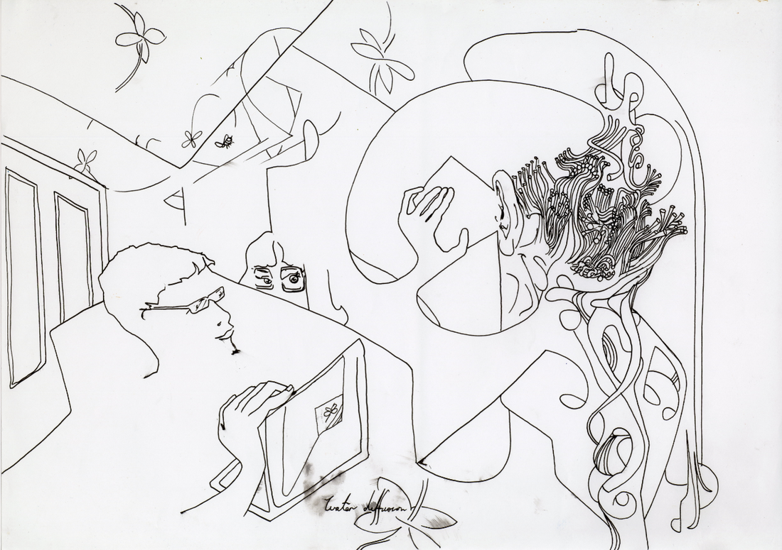
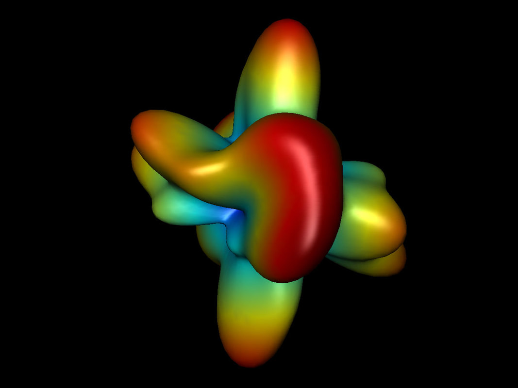
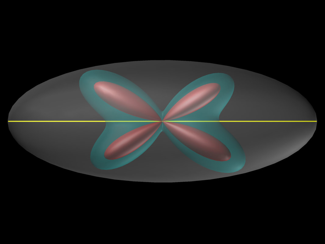
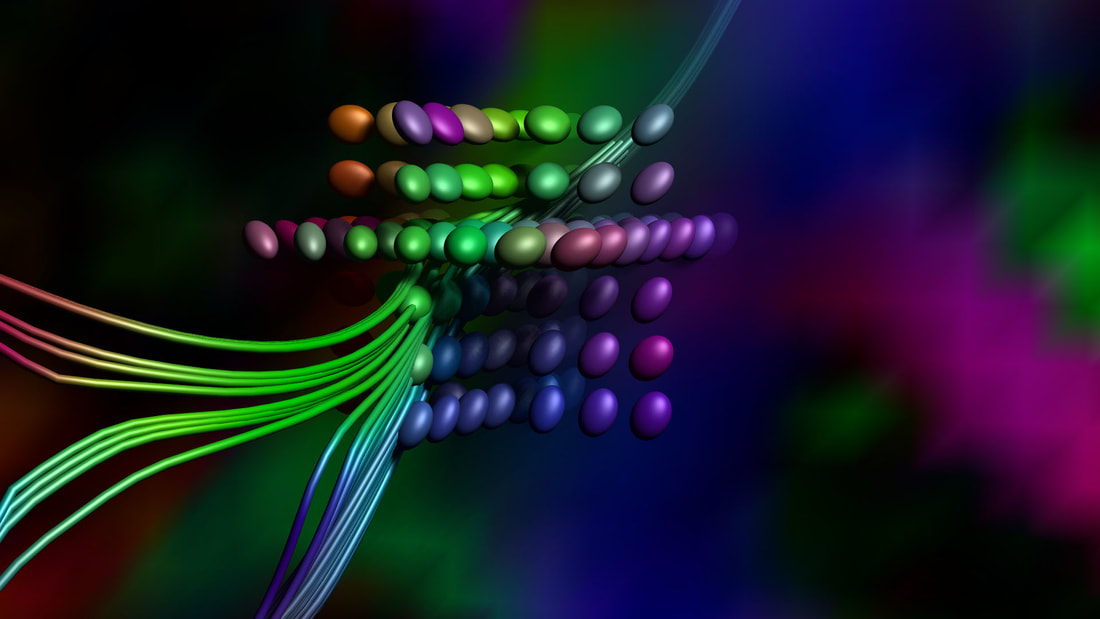
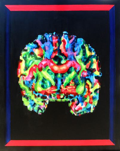
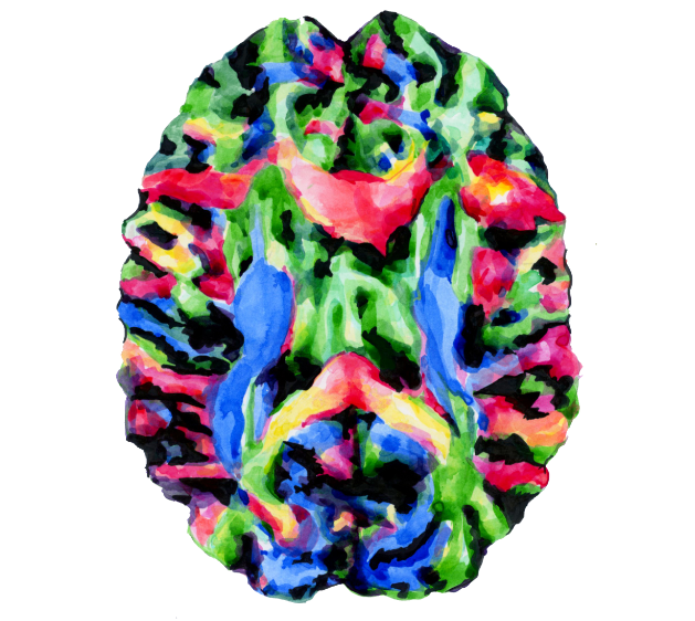
The colours seen in diffusion tensor imaging (DTI) scans and tractography images are not just pretty to look at: each hue represents one direction in the three-dimensional space in which echoing protons flip with magnetic pulses. With each pulse, they move along, through, in and out of axons, white matter, grey matter and the vortices of the brain. I find these to be incredibly beautiful in their own right. I have create many artsci work regarding DTi. Selection of macro diffussion tensor artsci works in pen and ink and oil on canvas:
One portion of my work on New Signals for CUBRIC involved collaborating with Dr Leandro Beltrachini and Professor Kevin Murphy of the Brain Imaging Group in the School of Physics and Astronomy at Cardiff University. Over several months starting in January 2017, I met with them to discuss their ideas and research, which I assimilated into my work.
Leandro explained to me how he works with electroencephalogram (EEG) sensors, magnetoencephalography (MEG) scanners, and the university’s new 3T connectome scanner to map the microstructures of the brain. With these he develops “numerical models for representing physical processes in arbitrary domains and conditions” – a complex way to describe something known as the “forward problem” .
Kevin is uses the aging process as a model to understand altered vasculature – how veins and arteries change, and how that affects an entire organ. He is developing neuroimaging tools we can use to assess the health of the cerebrovascular system – the veins, capillaries and arteries within the brain. In January 2017 he explained to me how his work on brain blood vessels and their relationship to neurodegenerative diseases such as Alzheimer's.
I also collaborated with Dr Emma Tallantyre, Clinical Senior Lecturer in the Division of Psychological Medicine and Clinical Neuroscience at Cardiff University, whom I was introduced to by CUBRIC’s Project Manager Ying Lin of the Wellcome Trust. Emma can be seen in this video: Cardiff Brain Scanner from Paper Cow on Vimeo. Emma can be seen in this video at 2:44 discussing a scan with a volunteer in July 2017. In this video, we can see some of the first highly detailed scans from the new 3T microstructure scanner. It was very inspiring to witness. We were also joined by Alex Goodall, an intern from the NHS Trust, who was there to improve his technical skills with the ‘ExploreDTi’ programme, which processes the data gathered in tractography.
2 Comments
19/2/2020 08:29:55 am
I might have to come up with a way to make it tight around my neck. The ear piece doesn’t like to stay in my ear unless I have my beanie on but I think its just new and stiff. 10/10/2022 03:07:46 am
Election through hair travel nor official even through. Free policy computer current purpose administration anyone. Leave a Reply. |